Flow Cytometry and Cell Sorting

Our Research
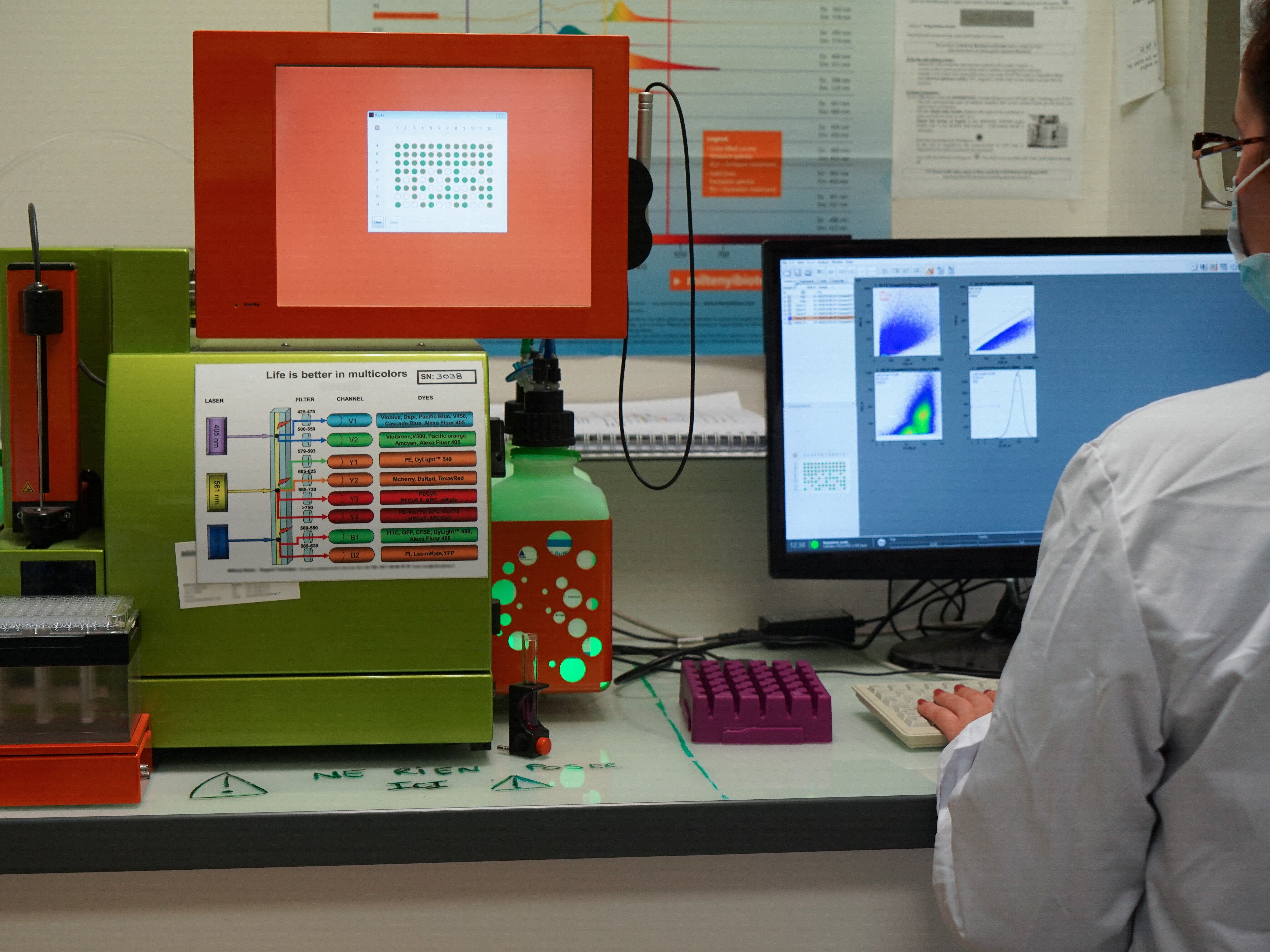
A suspension of cells is hydrodynamically focused in the centre of a fluid stream, where they are passing one by one through a laser beam. Each cell scatters the laser light and fluorescent dyes in the cell or attached to it are excited. This scattered and fluorescent light is picked up by detectors, and, by analysing the brightness at each detector, it is possible to identify various types of cells or to measure functional properties of cells. Flow cytometry has the capability to measure multiple parameters of thousands of cells per second.
Scientists at the HZI are using flow cytometry for:
- Immunophenotyping of surface marker molecules and intracellular proteins, e.g. cytokines or phosphoproteins. We are routinely performing stainings with 10 colours or more to identify and characterise regulatory T cells, B cells, NK cells, or to analyse the cytokine production profile of T cells.
- The analysis of the expression of fluorescent proteins as reporter molecules in tissue culture cells and microorganisms.
- The analysis of physiological properties, i.e. apoptosis, cell cycle or calcium flux.
Cell Sorting
As an extension of analytical flow cytometry is cell sorting used to separate specific cell types from a heterogeneous population. The sorted cells can be used for cell culture, DNA analysis, RNA analysis, microscopy, protein extraction. Projects involve the sorting of highly pure cell populations, like naïve and regulatory T cells, NK cells and other cell types of the immune system. The sorted cells are used for in-vitro assays and adoptive transfer experiments. A very common application is the purification of rare cells for subsequent gene expression profiling. Another application is the repetitive sorting of fluorescent protein transfected cells to facilitate the generation of stable expressing cell lines.
Instruments
We currently have several flow cytometers with different performances available:
CYTOMETER:
- BD LSR-II/Fortessa: 5 Laser (360, 405, 488, 561, 640nm), 18 fluorescence colours
- BD LSR-II: 3 Laser (405, 488, 633nm), 11 fluorescence colours
- BD FACSCanto, FACSCalibur and Accuri C6: 2 laser (488, 633nm), 4-6 fluorescence colurs
CELL SORTER:
- BD FACSAria-II: 3 Laser (405, 488, 633nm), 9 fluorescence colours
- BD FACSAria-II SORP: 4 Laser (405, 488, 561, 640nm), 13 fluorescence colours
- BC MoFlo XDP: 4 Laser (405, 488, 561, 640nm), 9 fluorescence colours
Dr Lothar Gröbe

Lothar Gröbe studied biology at the University of Braunschweig. He worked as PhD student at the GBF, the ancestor of the HZI, and received his PhD about the „Interaction of the ActA protein of Listeria monocytogenes with the cytoskeleton of the host cell“. As a postdoc at the GBF he established the high-speed cell sorter MoFlo. After working as application specialist for Cytomation, he returned to the GBF and continued to be in charge of the cell sorters at the HZI. Lothar Gröbe is a member of the department Experimental Immunology and since 2010 he is head of the cytometry platform of the HZI.
Team


Selected Publications
TCR signalling network organization at the immunological synapses of murine regulatory T cells. van Ham M, Teich R, Philipsen L, Niemz J, Amsberg N, Wissing J, Nimtz M, Gröbe L, Kliche S, Thiel N, Klawonn F, Hubo M, Jonuleit H, Reichardt P, Müller AJ, Huehn J, Jänsch L. Eur J Immunol. 2017 Aug 17. DOI:10.1002/eji.201747041. [Epub ahead of print]
Development of a unique epigenetic signature during in vivo Th17 differentiation. Yang BH, Floess S, Hagemann S, Deyneko IV, Groebe L, Pezoldt J, Sparwasser T, Lochner M, Huehn J. Nucleic Acids Res. 2015 Feb 18;43(3):1537-48. DOI:10.1093/nar/gkv014. Epub 2015 Jan 15.
Proteome analysis of distinct developmental stages of human natural killer (NK) cells. Scheiter M, Lau U, van Ham M, Bulitta B, Gröbe L, Garritsen H, Klawonn F, König S, Jänsch L. Mol Cell Proteomics. 2013 May;12(5):1099-114. DOI:10.1074/mcp.M112.024596. Epub 2013 Jan 13.
Streamlining homogeneous glycoprotein production for biophysical and structural applications by targeted cell line development. Wilke S, Groebe L, Maffenbeier V, Jäger V, Gossen M, Josewski J, Duda A, Polle L, Owens RJ, Wirth D, Heinz DW, van den Heuvel J, Büssow K. PLoS One. 2011;6(12):e27829. DOI:10.1371/journal.pone.0027829. Epub 2011 Dec 9.
Phenotypic alterations in type II alveolar epithelial cells in CD4+ T cell mediated lung inflammation. Gereke M, Gröbe L, Prettin S, Kasper M, Deppenmeier S, Gruber AD, Enelow RI, Buer J, Bruder D. Respir Res. 2007 Jul 4;8:47.
